- 首页
- 关于我们
概况介绍 研究方向 人才培养 委员会/committee
- 员工团队
特聘专家 主要领导 行政团队 技术团队 PI团队- 科研平台
中心实验室 实验动物中心 生物样本库- 最新资讯
- 首页
- 关于我们
概况介绍 研究方向 人才培养 委员会/committee
- 员工团队
特聘专家 主要领导 行政团队 技术团队 PI团队- 科研平台
中心实验室 实验动物中心 生物样本库- 最新资讯
Construction and characteristics of an adjustable biomechanical in vitro corneal stromal model simulating keratoconus pathological features发布日期: 2025-05-06
Abstract
Keratoconus (KC) is a prevalent ectatic corneal disease influenced by corneal biomechanics, though its pathogenesis remains unclear. Constructing an in vitro corneal model with adjustable biomechanics is essential for studying keratoconus. In this study, a KC disease group (L group) was created using a low matrix stiffness collagen hydrogel containing human corneal stromal cells (hCSCs), while a high matrix stiffness group (H group) was established using a plastic compression method. Additionally, a cross-linking treatment model was applied to the L group using corneal collagen cross-linking (CXL). Results showed that the matrix stiffness of the L group was significantly lower compared to the H group. Both the L group and clinical KC samples exhibited lower collagen fibrils density. Transcriptomic and proteomic analyses revealed decreased antioxidant capacity and increased levels of inflammatory factors and reactive oxygen species (ROS) in the L group. The NRF2 pathway activity was downregulated. In the L group cells, inflammation-related genes were upregulated, antioxidantrelated genes were downregulated, and ROS levels were elevated, accompanied by a reduction in mitochondrial membrane potential. The concentrations of IL-6 and TNF-α in the culture medium were increased. Histological results indicated that the L group, similar to clinical KC samples, exhibited high expression of inflammatory and oxidative stress factors, while signals of antioxidant-related factors were decreased. After cross-linking treatment, the L group demonstrated increased matrix stiffness, higher collagen fibrils density, upregulated expression of antioxidant factors, and decreased expression of inflammatory and immune factors. This study successfully established an in vitro corneal stromal model with adjustable matrix stiffness, providing a platform for investigating the pathogenesis of KC.
上一篇The influence of femtosecond laser intrastromal lenticules on the characteristics and maturity in tissue-engineered stem cellderived retinal pigment epithelium sheets下一篇ROS-Responsive Microneedle Patches Enable Peri-Lacrimal Gland Therapeutic Administration for Long-Acting Therapy of Sjögren’s Syndrome-Related Dry Eye相关文章-
近期,爱尔眼科长沙医学中心陈建苏教授、唐仕波教授、崔泽凯副教授团队在角膜损伤修复和视网膜疾病治疗方面研究取得了多项令人瞩目的成果,为了解致盲性角膜和视网膜疾病的...2025-08-04
-
AbstractKeratoconus (KC) is a prevalent ectatic corneal disease influenced by co...2025-05-06
-
Corneal nerve wounding often causes abnormalities in the cornea and even blindne...2024-09-04
-
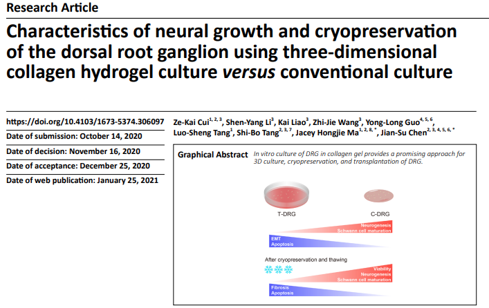
In vertebrates, most somatosensory pathways begin with the activation of dorsal ...2024-09-04
-
本申请涉及细胞和器官培养技术领域,公开了一种三维类器官灌流培养器具,包括:细胞培养室,细胞培养室用于盛放培养基;多个换液槽,设于细胞培养室,用于更换细胞培养室中...2024-09-04
-
Purpose: We performed a bioinformatic transcriptome analysis to determine the al...2024-09-03
- 员工团队
- 员工团队


