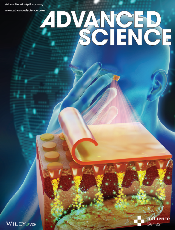
近日,爱尔眼科与南开大学联合科研团队取得重大突破,其研究成果在国际学术期刊《Advanced Science》(中科院 1区 TOP,影响因子14.3)上重磅发...
2025-05-19

Abstract
Previous
research has shown that CXCR5−/− mice develop retinal
degeneration (RD) with age, a characteristic related to age macular
degeneration (AMD). RD in these mice is not well-understood, and in this study,
we sought to characterize further the RD phenotype and to gain mechanistic insights
into the function of CXCR5 in the retina. CXCR5−/− and WT
control mice were used. Fundus images demonstrated a significant (p <
0.001) increase of hypo-pigmented spots in the retina of aged CXCR5−/− mice
compared with WT control mice. PAS staining indicated localization of deposits
in the sub-retinal pigment epithelia (RPE) layer. AMD-associated proteins
Cryab, amyloid beta, and C3d were detected within the RPE/sub-RPE tissues by
immunofluorescence (IF). In addition, western blot analysis of COX-2, Arg1, and
VEGF-a revealed an increase in the signaling of these molecules within the
RPE/choroid complex. Transmission electron microscopy (TEM) indicated a
drusen-like structure of sub-RPE deposits with an accumulation of vacuolated
cellular debris. Loss of photoreceptors was detected by peanut lectin staining
and was corroborated by a reduction in MAP2 signaling. Loss of blood-retinal
barrier integrity was demonstrated by a reduction of ZO-1 expression. Inflammatory
cells were detected in the sub-RPE space, with an increase in IBA-1 positive
microglia cells on the surface of the RPE. Mass spectrometry analysis of CXCR5−/− mouse
RPE/choroid proteins extracts, separated by SDS-page and incubated with
autologous serum, identified autoantibodies against AMD-associated proteins:
Cryaa, Cryab, and Anxa2. In vitro evaluations in BV-2 cell
culture indicated a significant increase in production of Arg-1 (p <
0.001) and COX-2 (p < 0.01) in the presence of anti-CXCR5
antibody when compared with Igg-treated control BV-2 cells stimulated with IL-4
and TNFα/IFNγ, respectively. Anti-CXCR5 antibody treatment without stimulating
agents did not affect Arg-1 and COX-2 expression; this suggests that CXCR5 may
have a regulatory role in microglia cells activation. These results indicate
that with age, CXCR5−/− mice develop RD characterized by
microglia dysfunction, increased production of CXCL13 in the RPE progressive
photoreceptor, neuronal loss, and sub-RPE deposition of cellular debris,
resulting in the production of immunogenic proteins and autoimmune-mediated RD.